 English
 French
 German
 Spanish
 Belgium
 Italian
 Brazil
 Chinese (Simplified)
Related Articles
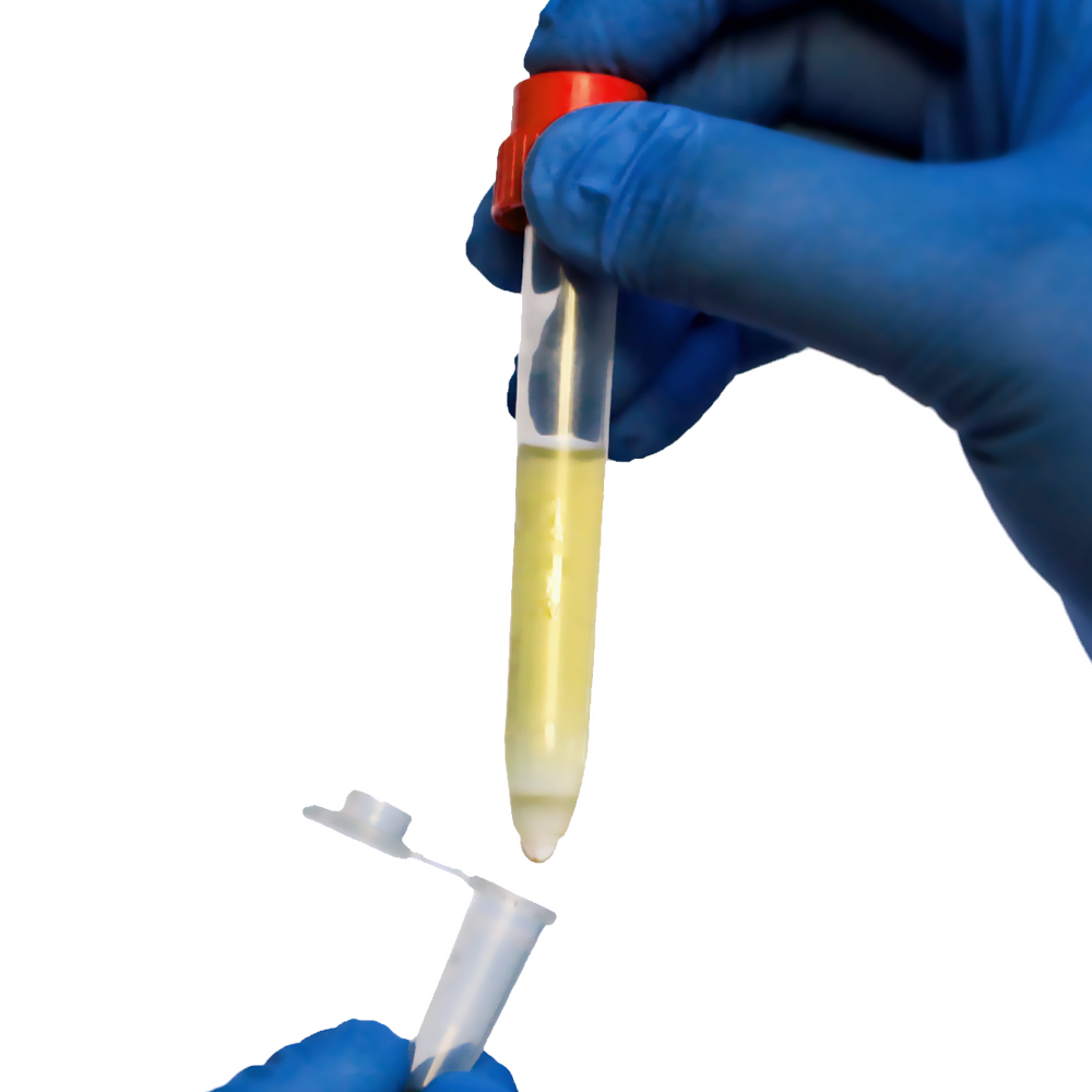
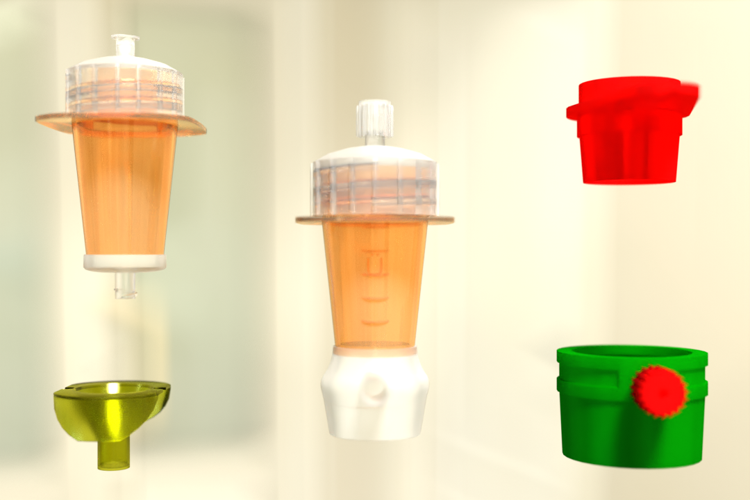
TwinSpin – A Device for Gravity Separation
TwinSpin centrifugation tubes pre-filled with Density Gradient Medium (DGM)
Why do we use it?
pluriStrainer PET vs. Nylon mesh – Which Material suits me? With our
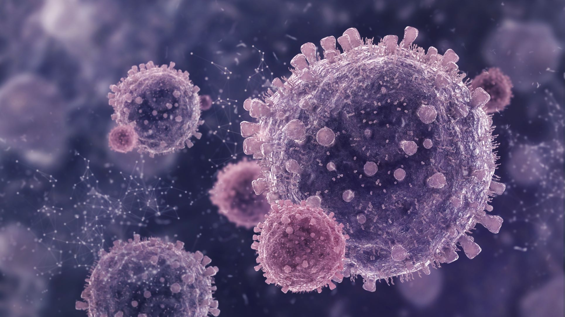
Circulating Tumor Cells (CTC)
Introduction Here we are presenting a new approach to colon carcinoma circu
Pluriselect’s All-Star-Strainer-Team: One Stop Solution to Particle Separation
PluriSelect developed a family of mesh-based sample preparation devices fo
A Detailed Guide on Positive Selection vs Negative Selection for Cell Isolation
Cells are an important research tool for studying various mechanisms in hea

T Cell Isolation:A Comprehensive Guide to the Key Components
T cells are a large and diverse group of lymphocytes that mature in the thy
What is Buffy Coat in Blood? Buffy Coat Preparation and Buffy Coat Cell Extraction
Buffy coat preparation is frequently the first step in further processing f
Necrosis Vs. Apoptosis: Necrotic Cell Death, Processes, & Apoptosis Steps
The stages of cell death differ depending on how a cell dies. When it comes