Showing 55–72 of 111 results
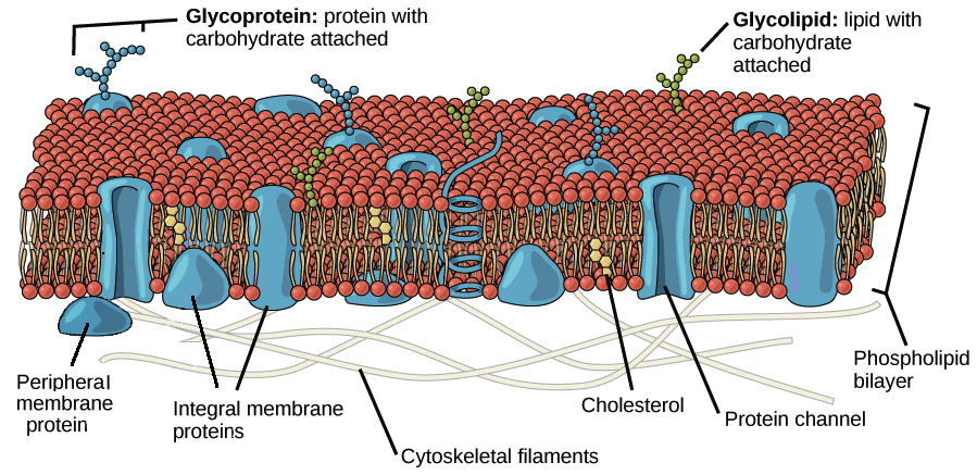
Image modified from OpenStax Biology: Source
Every cell is a particle BUT not every particle is a cell – there are differences in the separation tools!
Eukaryotic cells are surrounded by a flexible plasma membrane with specific surface structures (clusters of differentiation – CD’s)
(Image modified from OpenStax Biology https://www.khanacademy.org/science/high-school-biology/hs-cells/hs-the-cell-membrane/a/hs-the-cell-membrane-review)
The membrane structure explains the plasticity of the cell shape. In suspension they are spherical but in contact with a surface they change shape like an egg without the hard shell.
The bad News: The mesh-based separation of animal cells in suspension by size will only function by very big size differences (e.g.10 um from 80 um). Mesh openings are not holes in a membrane, they have an remarkable 3D structure due to the waeving process with fibers of a diameter of app. 30 um. Cells with their fluid membrane will change their shape and squeeze through much smaller openings as their ideal diameter.
The good news: The plasma membrane structure provides unique structures for a high specific cell separation, the surface molecules (CDs). All specific cell separation methods using specific anti-CD-monoclonal antibodies. Coupled on a solid support, they can be used for the positive cell separation (pluriBeads) or negative (untouched) cell separation or cell depletion (pluriSpin)
 English
 French
 German
 Spanish
 Belgium
 Italian
 Brazil
 Chinese (Simplified)
