Showing 73–90 of 111 results
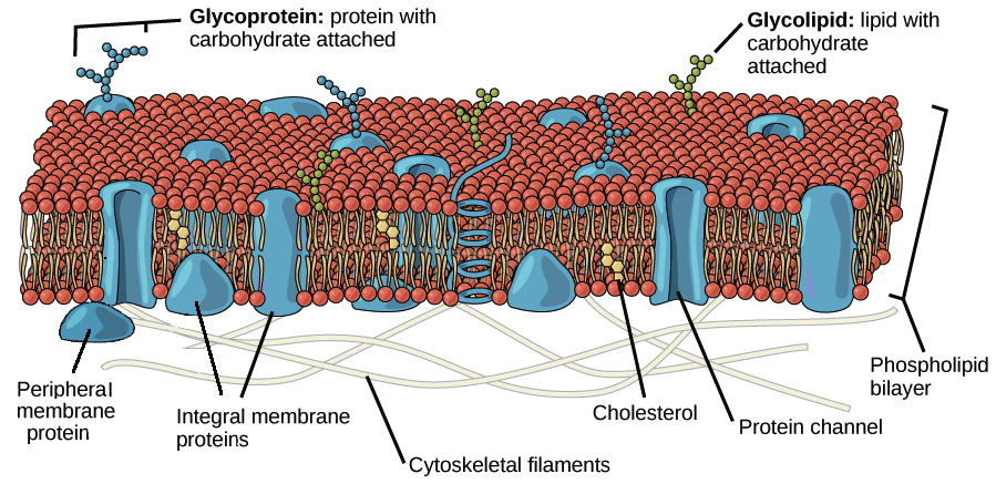
Image modified from OpenStax Biology: Source
Every cell is a particle BUT not every particle is a cell – there are differences in the separation tools!
Eukaryotic cells are surrounded by a flexible plasma membrane with specific surface structures (clusters of differentiation – CD’s)
(Image modified from OpenStax Biology https://www.khanacademy.org/science/high-school-biology/hs-cells/hs-the-cell-membrane/a/hs-the-cell-membrane-review)
The membrane structure explains the plasticity of the cell shape. In suspension they are spherical but in contact with a surface they change shape like an egg without the hard shell.
The bad News: The mesh-based separation of animal cells in suspension by size will only function by very big size differences (e.g.10 um from 80 um). Mesh openings are not holes in a membrane, they have an remarkable 3D structure due to the waeving process with fibers of a diameter of app. 30 um. Cells with their fluid membrane will change their shape and squeeze through much smaller openings as their ideal diameter.
The good news: The plasma membrane structure provides unique structures for a high specific cell separation, the surface molecules (CDs). All specific cell separation methods using specific anti-CD-monoclonal antibodies. Coupled on a solid support, they can be used for the positive cell separation (pluriBeads) or negative (untouched) cell separation or cell depletion (pluriSpin)
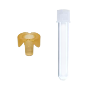
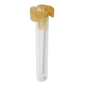
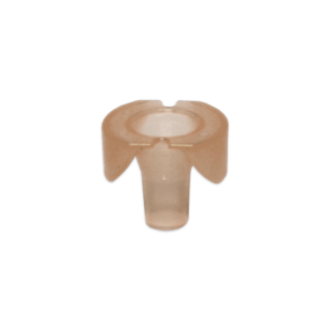
Bundle Ministrainer 100 µm mesh and Flow Cytometry Tubes, sterile
Price : NT$2,370.00 – NT$48,000.00
 English
 French
 German
 Spanish
 Belgium
 Italian
 Brazil
 Chinese (Simplified)
